
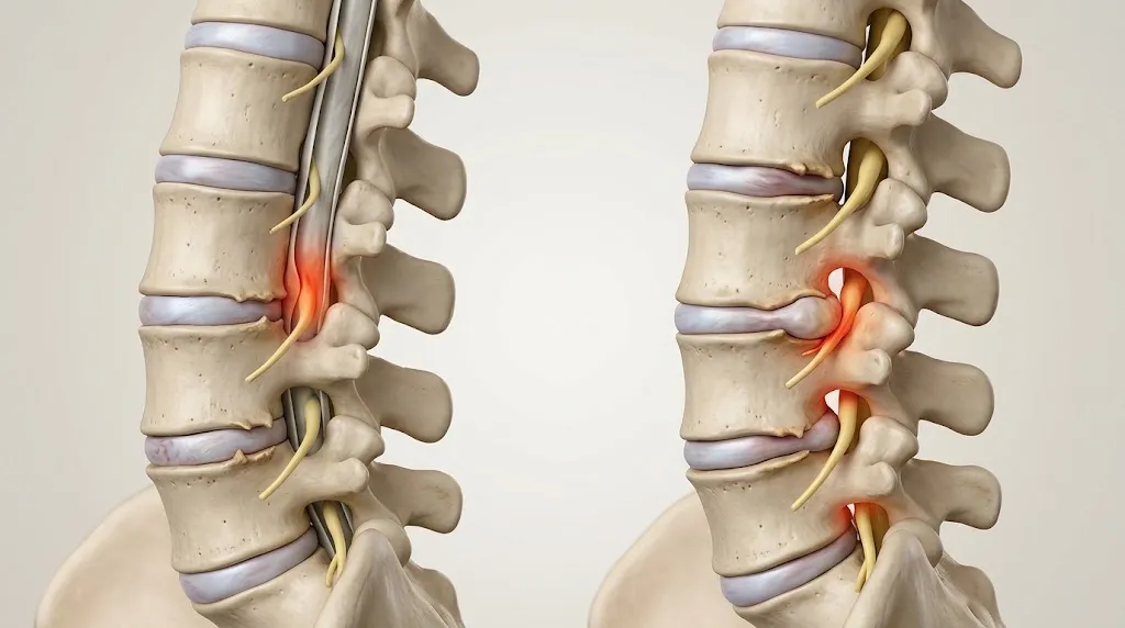
Which Is Worse: Spinal Stenosis or Foraminal Stenosis? 5 Key Differences
This article, “Which Is Worse: Spinal Stenosis or Foraminal Stenosis? 5 Key Differences,” was written by Dr. John Ma (Orthopedic Surgery).
Medical Disclaimer: The information provided in this article is for educational and informational purposes only. It is not intended to replace professional medical advice, diagnosis, or treatment. Spinal stenosis and foraminal stenosis can affect individuals differently, and diagnosis and treatment decisions should be made by a qualified healthcare professional based on individual symptoms and medical findings. Always consult your doctor or an appropriately qualified specialist about any health concerns. Seek urgent medical attention if you experience new loss of bladder or bowel control, numbness around the groin or saddle area, or rapidly worsening leg weakness.
A scan report that mentions both spinal stenosis and foraminal stenosis naturally raises the question of which is worse, spinal stenosis or foraminal stenosis. The short answer is that neither label settles it. Foraminal stenosis is one anatomical form of spinal stenosis, so these are not two unrelated diseases. How serious the problem is depends on where the narrowing sits, which nerve structures it presses on, how severe the pressure is, and whether it is causing symptoms. Many people have narrowing on imaging and never notice it.
This guide explains the anatomy, the five main differences, the symptoms that point to more serious compression, and how doctors diagnose and treat both conditions.
How foraminal stenosis fits inside spinal stenosis
Spinal stenosis means that spaces within the spine have become narrower, which can put pressure on the spinal cord, the cauda equina (the bundle of nerve roots in the lower back) or individual nerve roots. It can occur in the neck (cervical), mid-back (thoracic) or lower back (lumbar), and it is most common in the lumbar spine, followed by the neck.
Three areas matter when reading a report:
- The central spinal canal is the main passage for the spinal cord, and below it the cauda equina.
- The lateral recess is the side pocket of the canal where each nerve root travels before it leaves.
- The intervertebral foramen is the opening between two vertebrae through which a spinal nerve exits.
Narrowing can occur in any of these places, alone or in combination. Foraminal stenosis, which doctors also call neural foraminal narrowing, is narrowing at the exit. Cleveland Clinic describes it as a type of spinal stenosis that affects the neural foramen. clevelandclinic
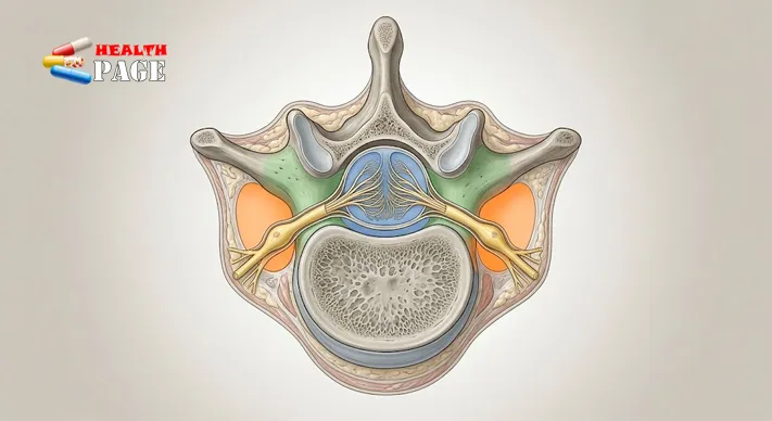
Spinal canal stenosis vs. foraminal stenosis: 5 key differences
The table gives a quick comparison, and the sections after it explain each row. This is also where the question of which is worse becomes easier to answer.
| Feature | Central (canal) stenosis | Foraminal stenosis |
|---|---|---|
| 1. Location | Main spinal canal | Nerve exit opening |
| 2. Structures affected | Spinal cord, cauda equina or several roots | Usually one exiting nerve root |
| 3. Typical symptoms | Walking-related pain, heaviness, neurogenic claudication | Radiating pain, tingling or weakness along one nerve |
| 4. Main concern | Cord or cauda equina compression in severe cases | Persistent radiculopathy, sensory loss, weakness |
| 5. Treatment focus | Decompression of the canal if needed | Relief of one root, sometimes by foraminotomy |
Difference 1: where the narrowing is. Central stenosis reduces the space in the main canal. Foraminal stenosis reduces the space in a side opening, so the problem is local to one exit point.
Difference 2: which structures are under pressure. In the neck, central narrowing can compress the spinal cord itself. In the lower back the cord has ended, so central narrowing affects the cauda equina or several roots at once. Foraminal narrowing more often affects the single root passing through that opening, on one side or both.
Difference 3: the pain pattern. Central stenosis in the lumbar spine is classically linked to neurogenic claudication, meaning leg pain, cramping or heaviness that builds with standing or walking and eases when sitting or leaning forward. This is because arching the back narrows the canal further. Foraminal stenosis tends to produce symptoms that follow one nerve’s path into an arm or leg.
Difference 4: severity and complications. This is the heart of the “which is worse” question, and the honest answer is that either can be serious. Severe cervical central stenosis can compress the spinal cord and cause cervical myelopathy. Severe lumbar central stenosis can, rarely, compress the cauda equina. Foraminal stenosis typically affects a single nerve root, but that can still mean significant pain, numbness or weakness in the limb. Cleveland Clinic also notes that only 17.5% of people with severe foraminal stenosis have symptoms, which shows why the degree of narrowing on a scan does not predict how a person feels.
Difference 5: treatment. Both are usually managed without surgery at first. If surgery is needed, the plan follows the location of the problem: decompression of the canal for central stenosis, or widening of the foramen for a trapped exiting root.
For anyone comparing central stenosis vs foraminal stenosis, the useful takeaway is that the more worrying features are those that suggest compression of the cord or cauda equina, or progressive weakness, whichever label appears on the report.
Lateral recess stenosis and other patterns
Lateral recess stenosis deserves a mention because it behaves much like foraminal stenosis. The recess is the passage inside the canal that leads to the exit, so pressure there can irritate the same root and produce similar leg or arm symptoms. Some reports also describe circumferential narrowing, where several dimensions of the canal are reduced together. This is why an MRI report may list central, lateral recess and foraminal findings separately, and why a clinician has to decide which one matches the person’s symptoms.
Foraminal stenosis symptoms
Foraminal stenosis symptoms depend on which nerve root is affected. Common features include:
- Radiating pain, which may feel sharp, burning or electric
- Tingling or pins and needles
- Numbness in part of an arm, hand, leg or foot
- Weakness in muscles supplied by that nerve
- Reduced reflexes or sensation found on examination
In the neck, a narrowed foramen can irritate roots supplying the shoulder, arm and hand. People may notice neck pain, pain travelling down one arm, fingertip tingling or a weaker grip. This pattern is called cervical radiculopathy. Foraminal narrowing from arthritic change is a common cause, and the same degenerative process can also produce shoulder-blade pain, as described in this guide to scapular pain caused by cervical spondylosis.
In the lower back, the symptoms often involve the buttock, leg and foot. Someone might have low back pain with leg pain on one side, tingling in the foot or a leg that tires on stairs. If you are trying to work out why you have numbness in one leg, a pinched lumbar nerve root is one of several possible explanations, though circulation problems and other nerve conditions need to be considered as well.
Pain that runs from the lower back down the leg is often called sciatica, and it is a symptom pattern rather than a diagnosis. Foraminal narrowing, lateral recess narrowing and a herniated disc can each produce it, which is one reason imaging has to be matched to the person’s examination.
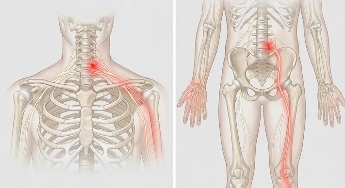
Severe spinal stenosis symptoms: warning signs
Most people with stenosis have a slow, manageable course. Certain changes suggest the nerves or spinal cord are under more serious pressure, and these are the severe spinal stenosis symptoms worth recognising early.
Signs that compression may be getting worse:
- Pain that is steadily increasing in the back, neck, arms or legs
- Numbness that spreads or deepens
- Weakness that progresses, such as a foot that begins to catch or drag
- A shrinking walking distance, or new unsteadiness
- Difficulty with everyday tasks
Possible cauda equina syndrome (emergency):
- New difficulty starting to urinate, or being unable to urinate
- Loss of bladder or bowel control
- Numbness around the groin, genitals or saddle area
- New or rapidly worsening weakness in one or both legs
These symptoms need emergency assessment the same day. They should not wait for a routine appointment. NHS patient information for lumbar stenosis describes cauda equina syndrome and spinal myelopathy as rare conditions where the spinal cord gets compressed, which is accurate but should not delay care if the signs appear. sfh-tr
Possible cervical myelopathy (needs prompt specialist review): clumsy hands, trouble with buttons or handwriting, stiffness or weakness in the arms or legs, and poor balance.
Compression of the spinal cord is a different problem from compression of a single nerve root. Both can be serious, but they produce different symptoms and call for different levels of urgency.
What causes spinal and foraminal stenosis?
Most stenosis is acquired through gradual wear. As discs lose height, the vertebrae move closer together and the foramina get smaller. Degenerative disc changes are therefore one of the most common routes to foraminal narrowing.
Other contributors include bone spurs (osteophytes), enlarged facet joints, thickening of the ligamentum flavum, a vertebra slipping forward over another (degenerative spondylolisthesis), and past spinal injury. Some people are born with a naturally narrow canal and have less margin for age-related change. Congenital and degenerative factors can also combine.
Disc terminology often appears on reports too. Bulge, protrusion, extrusion and sequestration describe the shape of disc material rather than stages on a fixed timetable, and a bulge does not necessarily progress to the others. A disc abnormality also does not automatically mean symptomatic stenosis.
How doctors diagnose the condition
Diagnosis starts with the story: where the pain is, whether it travels, what makes it better or worse, and whether walking, standing or leaning forward changes it. A neurological examination then checks strength, sensation, reflexes, balance and gait.
MRI is usually the preferred scan because it shows the discs, nerve roots, ligaments and the extent of any compression. Radiologists use different sequences to highlight different tissues, and this explainer on how T1 and T2 MRI sequences differ helps make sense of the images. CT shows bone detail such as spurs and facet changes and can be paired with myelography in selected cases. X-rays reveal alignment, disc-space narrowing and slippage but are less useful for nerve compression.
The most important point is that images must be read alongside symptoms and examination. Measurements such as canal diameter or foraminal grading are useful reference points, not universal cutoffs. Some people have severe-looking narrowing and few symptoms, while others have moderate narrowing and considerable nerve irritation. Having several levels of narrowing is common, and the clinician’s job is to decide which finding matches the complaint.
Central and foraminal stenosis can also coexist. A scan might show central narrowing at L4–L5 with right-sided foraminal narrowing at the same level and further foraminal change at L5–S1.
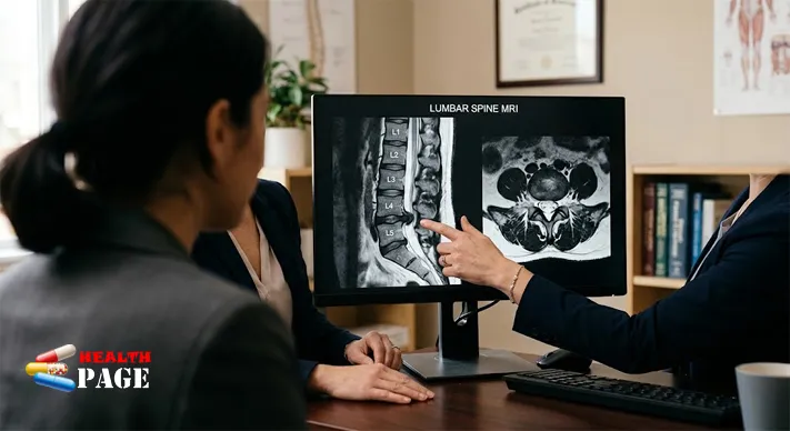
Treatment options
Treatment is individual, and narrowing on a scan alone is not a reason for surgery.
Non-surgical care is the usual starting point when there is no severe or progressive neurological problem. Physical therapy can improve flexibility, core strength, balance and walking tolerance, with exercises chosen to suit the affected region. Activity changes can help too; people with lumbar stenosis often find that sitting or leaning forward gives relief. Anti-inflammatory medicines and nerve-pain medicines may be used depending on health history and other drugs. NHS patient information notes that in most cases symptoms can be managed with simple treatment that does not require an operation. sfh-tr
Epidural steroid injections may calm radiating pain for a time, but they do not remove bone spurs or permanently widen a narrowed space. The NIAMS summary of the LESS trial found that adding corticosteroid to numbing medication gave only a small benefit over the numbing medication alone, so expectations should be realistic. nih
Surgery is considered when symptoms stay disabling despite appropriate non-surgical treatment, when weakness progresses, or when there is myelopathy or another serious neurological complication. One NHS patient leaflet lists no improvement with conservative treatment in three to six months as one situation where an operation may be discussed. In the large SPORT trial summarised by NIAMS, surgery was more effective than nonsurgical treatment in relieving symptoms and improving function for people with spinal stenosis, although individual results vary and surgery carries its own risks. sfh-trnih
Common procedures include:
- Laminectomy or laminotomy: removing all or part of the lamina to make more room in the canal
- Foraminotomy: enlarging the foramen to free an exiting nerve root
- Spinal fusion: added when there is instability or slippage, using bone graft and usually instrumentation
Minimally invasive techniques such as unilateral biportal endoscopy (UBE), which uses one small portal for the camera and another for instruments, are used by some surgeons for selected lumbar decompressions. Suitability depends on the person’s anatomy, the diagnosis and the surgeon’s experience, and no technique is better for everyone.
After decompression, the removed tissue does not simply return, but degeneration can continue. New narrowing or disc problems can develop later, including at neighbouring levels, so following the surgeon’s rehabilitation advice matters.
Seek medical assessment for persistent back or neck pain, pain that spreads into an arm or leg, recurrent numbness, weakness, or a change in balance or walking. For a broader look at bone and joint topics, HealthPage’s orthopedics guides cover related conditions and recovery.
FAQs
Is foraminal stenosis serious?
Often it is not. Many people never develop symptoms, and when they do, the nerve root frequently settles with non-surgical care. It becomes more concerning when weakness progresses or pain does not improve with treatment.
Can foraminal stenosis go away on its own?
The structural narrowing usually does not reverse, but symptoms often improve as inflammation around the nerve calms. A person who is getting worse rather than better should be reassessed.
Can spinal stenosis cause paralysis?
Severe compression of the spinal cord or nerves can cause major neurological loss, but this is not the typical outcome. Progressive weakness, loss of bladder or bowel control, or saddle numbness need urgent evaluation.
Can you have both central and foraminal stenosis?
Yes. They can occur at the same level or at different levels, and a clinician has to work out which finding explains the symptoms.
Which scan is best?
MRI is commonly preferred for nerves, discs and the canal, while CT adds detail about bone. The choice depends on the question being asked and the person’s circumstances.
Conclusion
So, which is worse, spinal stenosis or foraminal stenosis? Neither is automatically worse, because foraminal stenosis is part of the spinal stenosis family. What matters is the affected structure and the symptoms: cord or cauda equina compression, progressive weakness and bladder or bowel changes carry more urgency than stable, one-sided nerve irritation. If you have pain, numbness or weakness that is not settling, a clinical assessment that links your symptoms to your imaging is the most useful next step, and any red-flag symptom should be treated as an emergency.
This article is for general information and is not a substitute for personal medical advice.
References
- Cleveland Clinic. “Foraminal Stenosis.” https://my.clevelandclinic.org/health/diseases/24856-foraminal-stenosis
- Sherwood Forest Hospitals NHS Foundation Trust. “Lumbar spine stenosis” (patient information). https://sfh-tr.nhs.uk/patients-and-visiting/patient-information-leaflets/musculoskeletal-msk/lumbar-spine-stenosis/
- National Institute of Arthritis and Musculoskeletal and Skin Diseases (NIAMS), NIH. “Spinal Stenosis: Research & Resources.” https://www.niams.nih.gov/index.php/health-topics/spinal-stenosis/more-info

Dr. John Ma is a dedicated Orthopaedic Surgeon with extensive experience in musculoskeletal health and surgical care. He currently serves at Royal Brisbane and Women’s Hospital, Australia, where he provides expert treatment for complex orthopaedic conditions. Passionate about advancing patient outcomes, Dr. Ma combines evidence‑based practice with compassionate care, helping individuals restore mobility and improve quality of life. Through his contributions to healthcare, he continues to support patients in achieving long‑term wellness and recovery.