
How Does the Body Make Urine? 5 Steps of Urine Production Explained
Medical Disclaimer: This article is intended for educational and informational purposes only. It explains how the human body produces urine and how the kidneys regulate fluid balance and remove waste. It is not a substitute for professional medical advice, diagnosis, or treatment. Always consult a qualified healthcare professional if you have concerns about changes in urination, urine color, or kidney health.
How does the body make urine? The kidneys filter blood, take back almost everything useful from the filtered fluid and add a few extra wastes. The leftover fluid passes through the ureters to the bladder, where it is stored until you urinate.
It is easy to picture the kidneys as strainers that catch waste and let everything else flow on. They do something more discerning. Each day they filter far more fluid than the body could afford to lose, then reclaim nearly all of it. The final urine reflects a series of decisions about what to keep and what to release. This article follows that process in five steps and introduces the structures involved. It also covers what changes urine volume and color, and when a change in urination deserves a medical opinion.
The organs that make and move urine
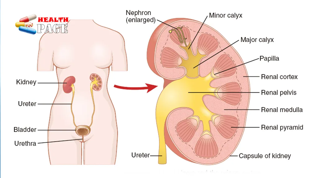
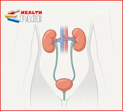
Four organs share the work. The two kidneys are bean-shaped, about the size of a fist, and sit just below the rib cage on either side of the spine. Besides making urine, they help balance water and minerals and regulate blood pressure and blood acidity. They also release erythropoietin, a hormone that supports red blood cell production.
Two narrow, muscular tubes called ureters carry urine from the kidneys to the bladder, pushing it along with rhythmic contractions known as peristalsis. The bladder is a hollow muscular organ. Its wall, the detrusor muscle, relaxes while urine collects and contracts when it is time to empty. The urethra then carries urine out of the body, with sphincter muscles acting as valves that decide when it is released.
The nephron: where urine is actually made
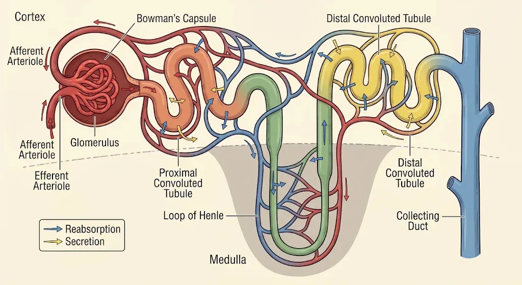
Each kidney contains roughly a million nephrons, the microscopic filtering units described in NIDDK’s overview of how the kidneys work. The exact number varies between people. A nephron has two main parts. The renal corpuscle is a tuft of capillaries called the glomerulus, wrapped in a cup-shaped Bowman’s capsule, and it is where filtration begins. The renal tubule that follows is a long, winding tube with distinct segments: the proximal convoluted tubule, the loop of Henle and the distal convoluted tubule. Several nephrons drain into a shared collecting duct, which helps set how concentrated the urine finally is.
How is urine produced in the kidneys? The five steps
The five steps below are an editorial way of organizing the journey. Physiologists usually describe urine formation as three processes: filtration, reabsorption and secretion. Steps 2 to 4 cover those, with the blood supply before them and the drainage afterward.
Step 1: Blood reaches the glomeruli
Blood enters each kidney through the renal artery, which divides into smaller and smaller vessels until it reaches the nephrons. Within a nephron, blood flows into the glomerulus through a tiny vessel called the afferent arteriole and exits through the efferent arteriole. The outflow vessel is narrower than the inflow vessel, which helps keep pressure high inside the glomerular capillaries. That pressure drives the next step. The kidneys receive roughly a fifth of the heart’s output at rest, so the body’s blood passes through them many times a day, giving them constant feedback on its composition.
Step 2: Glomerular filtration produces primary urine
How do kidneys filter blood in practice? Pressure inside the glomerular capillaries pushes water and small dissolved substances through a three-layered filtration barrier into Bowman’s capsule. The barrier consists of the capillary wall, a basement membrane and specialized cells called podocytes. The fluid collected is the glomerular filtrate, often called primary urine. It resembles blood plasma without the cells and large proteins, and it contains water, glucose, amino acids, sodium and other electrolytes, urea and uric acid.
Blood cells and most large proteins stay in the bloodstream because they cannot normally pass through. When the barrier is damaged, protein can leak into the urine. Short-lived rises after a fever or hard exercise are also common, as this explanation of a high urine protein result points out.
The volume involved is striking. Filtration runs at about 125 milliliters per minute in a healthy adult, a measure called the glomerular filtration rate (GFR). That works out to roughly 180 liters a day in OpenStax’s physiology text. GFR varies with body size, age and health, so a range of 150 to 180 liters is a reasonable ballpark. Almost none of it leaves the body.
Step 3: Tubular reabsorption returns useful substances to the blood
Having filtered a huge volume, the kidney now reverses most of it. Filtrate moves into the renal tubule, and the cells lining it move useful substances back into the surrounding capillaries. Think of a sorting line at a recycling plant: everything is pulled off the belt first, and the valuable items are picked out afterward.
Most of this happens in the proximal convoluted tubule, which reclaims roughly 65 to 70 percent of the filtered sodium and water. Water follows sodium by osmosis. Glucose and amino acids are normally almost completely reabsorbed there, provided blood levels and kidney function are normal. Chloride, bicarbonate and calcium also return in amounts that match the body’s needs. When blood glucose climbs very high, as in poorly controlled diabetes, the transport machinery becomes saturated and glucose spills into the urine. Without reabsorption, dehydration would set in within hours.
Step 4: Tubular secretion and urine concentration produce final urine
Reabsorption moves substances out of the tubule. Tubular secretion does the opposite, moving certain substances from the blood into the tubular fluid. It lets the kidneys remove hydrogen ions, which helps regulate acid-base balance. It also removes potassium when levels are high, along with many medicines and their breakdown products. Creatinine, a waste product of normal muscle metabolism, is mostly filtered, with a smaller portion added by secretion.
Meanwhile, the loop of Henle sets up the concentration of the urine. Its descending limb lets water leave the fluid, making it more concentrated. The ascending limb pumps out sodium and chloride but is largely impermeable to water, so the fluid becomes more dilute. The thick ascending segment alone reclaims about a quarter of the filtered sodium. Together, these movements build a salty gradient in the kidney’s medulla, which the collecting ducts use to draw water back when the body needs to conserve it.
Two hormones fine-tune the result. Antidiuretic hormone (ADH) makes the collecting ducts more permeable to water by adding aquaporin-2 water channels. More water then returns to the blood, and the urine becomes more concentrated. Aldosterone increases sodium reabsorption and potassium secretion in the distal nephron, with water balance affected indirectly. Under typical conditions, about 99 percent of the filtered water has been reclaimed by the end of this stage. What remains is final urine.
Step 5: Urine travels to the bladder and leaves the body
How does urine travel to the bladder from there? The route is a one-way drainage system:
- Collecting ducts carry urine toward the renal papillae.
- The papillae empty into the minor calyces, which merge into the major calyces.
- The renal pelvis collects urine from the calyces.
- Each ureter carries urine down to the bladder.
- The bladder stores it until urination, and the urethra carries it out of the body.
Peristaltic waves move urine down each ureter in small spurts rather than a steady stream. That is one reason a stone lodged in a ureter can be so painful, and this guide to common kidney stone symptoms explains what to watch for.
The bladder expands as urine accumulates, and stretch receptors in its wall signal the nervous system as it fills. When the moment is right, the brain permits the urination reflex. The detrusor muscle contracts, the sphincters relax and urine passes through the urethra. The external urethral sphincter is under voluntary control, which is why most adults can wait. The sensation of fullness varies between people and situations, so there is no single volume at which everyone feels the urge.
Urine production and urination are separate events. The kidneys make urine around the clock, and the bladder simply stores it until you go.
Primary urine versus final urine
This comparison explains why the kidneys can filter so much fluid and still produce so little urine.
| Feature | Primary urine (filtrate) | Final urine |
|---|---|---|
| How it forms | Glomerular filtration | Filtrate after reabsorption and secretion |
| Where it is found | Bowman’s capsule, start of the tubule | Collecting ducts onward |
| Daily volume | Roughly 150–180 L | Usually about 1–2 L |
| Glucose | Present | Normally absent |
| Amino acids | Present | Normally absent or very low |
| Urea | Present | More concentrated |
| Blood cells, large proteins | Normally absent | Normally absent |
The figure of about 99 percent water reabsorption applies under typical conditions. It shifts with hydration, hormones and health.
Why urine production is about more than waste removal
Urine formation is the kidneys’ main way of keeping the blood’s composition steady, a state called homeostasis. Four jobs stand out.
Water balance. When you are short of fluid, ADH levels generally rise, the collecting ducts reclaim more water, and urine becomes scarcer and darker. After a large drink, ADH generally falls and the surplus leaves as dilute urine.
Electrolytes. Sodium, potassium, chloride, calcium and phosphate are adjusted constantly. These minerals matter for nerve signaling, muscle contraction and the distribution of fluid in the body.
Acid-base balance. By secreting hydrogen ions, reabsorbing filtered bicarbonate and making new bicarbonate when needed, the kidneys help keep blood pH in a narrow range. The lungs work alongside them by regulating carbon dioxide.
Blood pressure. Through sodium and fluid handling, and through the release of renin, an enzyme that starts the renin-angiotensin-aldosterone system, the kidneys influence blood pressure.
What changes how much urine you make
No single volume is right for everyone. Many healthy adults pass roughly 1 to 2 liters a day, but body size, activity, climate, diet and health all move that number.
| Factor | Typical effect on urine |
|---|---|
| Fluid intake | More fluid generally means more urine |
| Dehydration or heavy sweating | Usually less, more concentrated urine |
| ADH | Raises water reabsorption and concentrates urine |
| Caffeine | Mild diuretic effect, mainly in people not used to it |
| Alcohol | Can suppress ADH and increase urine output |
| Diuretic medicines | Increase urine production |
| Health conditions | Kidney, heart and hormonal disorders can raise or lower output |
What urine contains and why its color changes
Urine is roughly 95 percent water. The rest is dissolved substances the body is clearing: urea from protein metabolism, creatinine from muscle metabolism, uric acid from the breakdown of purines, and electrolytes such as sodium, potassium and chloride. Smaller amounts of ammonium, phosphate and sulfate are also present. The mix shifts with hydration, diet, metabolism, hormones and kidney function.
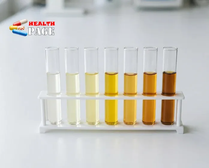
Color gives a rough, imperfect clue:
- Pale yellow: often relatively dilute urine.
- Dark yellow or amber: can mean concentrated urine, sometimes from too little fluid.
- Orange: may relate to certain medications, foods or medical conditions.
- Pink or red: can come from foods or medications, but may also be blood.
- Brown or tea-colored: has several possible causes, including dehydration, liver problems or muscle injury.
Pregnancy can add its own shifts, covered in this look at how urine color can change in early pregnancy. Color alone cannot reliably show hydration status or diagnose a disease. Occasional changes are often harmless.
When changes in urination deserve attention
Many urinary changes are temporary and harmless, but these are worth raising with a doctor:
- Pain or burning when urinating
- Blood in the urine
- Persistently dark or cloudy urine
- A marked drop in urine output
- Frequent urination, especially with excessive thirst
- Difficulty starting urination or emptying the bladder
- Persistently foamy urine
- Side or back pain with a fever
These symptoms have different causes and do not automatically point to kidney disease. Blood in the urine, for example, is most often caused by a urinary infection, and it can look pink, red or brown, according to Kidney Research UK. Kidney stones and other conditions can also cause it. A simple urine test is usually the first step in finding the cause, and a guide to what a urinalysis report measures shows what such a test looks at.
Seek prompt medical attention for visible blood in the urine, an inability to urinate, or markedly reduced urine output. This applies especially when swelling, breathlessness or feeling seriously unwell comes with it.
Frequently asked questions
How much urine do the kidneys make in a day?
Many healthy adults produce roughly 1 to 2 liters. Fluid intake, heat, exercise, diet and health can shift that figure considerably, so a different number is not automatically a problem.
Do the kidneys make urine all the time?
Yes. Filtration continues day and night. Output is often lower overnight, partly because ADH release tends to rise in the evening, so many people can sleep without waking to urinate.
Why do the kidneys reclaim so much water?
The filtered volume far exceeds what the body could replace. Without reabsorption, fluid would be lost faster than anyone could drink. Reclaiming it keeps blood volume, salt concentrations and hydration stable, and ADH lets the kidneys adjust how much comes back.
What is GFR, and why do doctors measure it?
GFR is the volume of filtrate both kidneys form each minute. Doctors usually estimate it (eGFR) from a blood creatinine result, adjusted for factors such as age and sex. It is one of the main indicators of kidney function, and results are read alongside urine tests and symptoms.
Why is my urine dark yellow?
Most often because it is concentrated, which happens when the kidneys conserve water. Foods, vitamins, medicines and some health conditions can also deepen the color. If it persists despite adequate fluids, or comes with other symptoms, speak with a healthcare professional.
Conclusion
Put simply, how does the body make urine? Blood reaches the glomeruli, which filter off a plasma-like fluid. The tubules then take back what the body needs, secrete extra wastes and adjust water and salt under hormonal control. The leftover fluid drains through the calyces and ureters to the bladder before leaving through the urethra. The kidneys act as selective regulators rather than simple filters, which is why urine volume and color shift with hydration, diet and health.
A practical next step is to notice your own patterns. Pale yellow urine on most days is typical for many people. Mention persistent changes, blood, pain or a fall in output to a healthcare professional rather than waiting for them to settle.
References
- National Institute of Diabetes and Digestive and Kidney Diseases (NIDDK), “Your Kidneys & How They Work”: https://www.niddk.nih.gov/health-information/kidney-disease/kidneys-how-they-work
- OpenStax, Anatomy and Physiology, “25.5 Physiology of Urine Formation”: https://openstax.org/books/anatomy-and-physiology/pages/25-5-physiology-of-urine-formation
- Kidney Research UK, “Blood in urine (haematuria): causes, symptoms and treatment”: https://www.kidneyresearchuk.org/conditions-symptoms/blood-in-urine/

Medical Reviewer: Dr. He Feng, MD
Specialist in Urology & Andrology | Clinical Contributor
Dr. He Feng is an expert urologist and andrologist practicing at
Beijing Jishuitan Hospital, widely recognized as the
Fourth Medical College of Peking University in China. With extensive clinical experience
in advanced genitourinary health, Dr. Feng specializes in the diagnostic assessment, management,
and treatment of complex urological conditions, obstructive urinary disorders, and male reproductive health.
His medical contributions on HealthPage focus on translating complex urological pathologies—such
as prostate health, urolithiasis (kidney stones), and renal system care—into highly accessible,
evidence-based guidance for patients globally.
Clinical Affiliation & Triage:
Beijing Jishuitan Hospital (The Fourth Medical College of Peking University)
Professional Inquiries:
[email protected]