
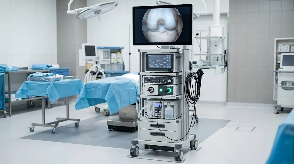
Arthroscopy Tower: Components, Uses, and How It Works
Medical Disclaimer: The article titled “Arthroscopy Tower: Components, Uses, and How It Works,” written by Dr. John Ma (Orthopedic Surgery), is provided for informational and educational purposes only. It is not intended to serve as a substitute for professional medical advice, diagnosis, or treatment. Always seek the advice of your physician or other qualified health provider with any questions you may have regarding a medical condition.
When a surgeon looks inside a knee or shoulder through an incision only a few millimetres wide, the view appears on a monitor across the room. The equipment behind that image is the arthroscopy tower, a mobile equipment trolley that brings together the camera system, light source, display and, in many configurations, fluid-management and powered-instrument controls used in minimally invasive joint surgery.
This article is written for medical students, healthcare trainees, operating-theatre staff and anyone curious about orthopaedic surgical equipment. It explains how the technology works. It is not a guide for patients preparing for their own procedure, and it is not training in how to operate medical devices, which always requires manufacturer instruction and supervised clinical practice. If you are facing arthroscopy yourself, your surgical team is the best source for what to expect, and the NHS arthroscopy overview is a good plain-language starting point.
How Does an Arthroscopy Tower Work?
An arthroscopy tower does not perform surgery. It produces, processes and displays the image that guides the surgeon, and it can support the instruments used inside the joint. Think of it as the surgical team’s shared window and utility cabinet in one.
The process follows a simple chain. A light source sends illumination through a fibre-optic cable into the arthroscope, which carries it into the joint. The arthroscope then relays the illuminated view back to a camera head. The camera converts that view into an electronic signal and passes it to a camera control unit, which processes it and sends the finished picture to a medical monitor. Meanwhile, an irrigation pump keeps sterile fluid moving through the joint so the view stays clear.
A distinction that confuses many newcomers is the difference between three terms that are often used loosely:
- The arthroscope is the slim optical instrument inserted into the joint.
- The arthroscopy camera system is the camera head, its cable and the processing unit that turns the optical view into a digital image.
- The arthroscopy tower is the whole supporting platform that houses the camera system, light source, monitor and other consoles.
So an arthroscope can exist without a tower in a teaching model or a bench test, but in an operating theatre it relies on the tower to be useful.
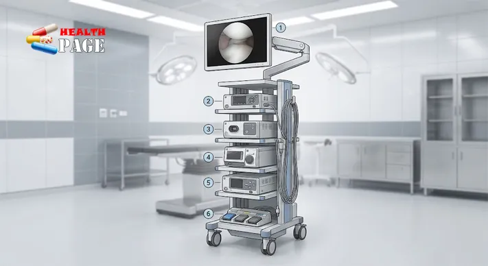
Main Arthroscopy Tower Components and Their Functions
Arthroscopy tower components vary by manufacturer and by what a hospital needs, so no single list fits every tower. Some modern units combine the camera processor, light source and image-recording functions into a single console, while older or more modular arrangements keep them separate. What follows is the common set of parts.
| Component | Main function | Usually essential? |
|---|---|---|
| Arthroscope | Optical instrument that gives the view inside the joint | Yes |
| Camera head | Captures the image from the arthroscope | Yes |
| Camera control unit (CCU) | Processes the signal and controls image settings | Yes |
| Light source | Provides illumination through a fibre-optic cable | Yes |
| Medical monitor | Displays the live image for the team | Yes |
| Irrigation pump | Circulates sterile fluid to keep the view clear | Common |
| Shaver system | Powered instrument for removing or smoothing tissue | Procedure-dependent |
| RF or ablation system | Delivers energy for selected tissue treatment | Procedure-dependent |
| Recording system | Saves images or video for documentation and teaching | Optional |
| Equipment trolley | Holds consoles and organises cables | Yes |
The imaging core
The arthroscope is the part that enters the joint. Its diameter depends on the joint involved, and smaller scopes are used in small spaces such as the wrist or ankle. The camera head attaches to the eyepiece end, and the control unit adjusts things such as brightness and colour balance. The light source matters more than it might seem: a joint is a closed, dark space, and image quality depends heavily on how well it is lit. Many current systems use LED light sources, though other types exist.
Fluid management and powered instruments
The irrigation pump is easy to overlook, yet it directly affects what the surgeon can see. The joint is distended with sterile fluid, which creates working space and washes away blood and debris. Pressure and flow are managed by the surgical team, and this is not something a general article can or should prescribe numerically, since appropriate settings depend on the joint, the procedure and the patient.
Shaver and ablation systems sit on the tower as separate control units or as modules that connect to handpieces and foot pedals. A shaver trims or smooths selected tissue, and radiofrequency devices can treat tissue using energy. Not every procedure needs both.
Do all towers contain the same equipment?
They do not. A basic diagnostic setup may need only an imaging chain and fluid supply, whereas a complex shoulder repair may call for powered instruments, ablation and a recording unit. Compatibility also matters: cables, scopes and handpieces are generally designed to work with specific systems, so mixing manufacturers’ parts without checking is not a safe assumption.
How an Arthroscopy Camera System Produces Images
The arthroscopy camera system is where the optical view becomes a digital one. Light reflected from the inside of the joint travels up the arthroscope’s lens system to the camera head. There, an image sensor converts the light into an electronic signal, much as a digital camera does, although in a sterile, tightly sealed housing built for repeated cleaning.
The camera control unit then handles the signal. It can adjust brightness, contrast and colour, and it passes the processed image to the monitor in real time. Many current systems offer high-definition or 4K output, which gives surgeons finer detail of cartilage, ligament and meniscus. Some towers also add features such as image capture or video recording for the patient record, which should follow the hospital’s privacy and consent policies.
A practical point for trainees: image quality is a chain, and weak links show up everywhere downstream. A fogged lens, a loose cable, a poorly matched light cable or a mis-set white balance can each make the picture worse, even when the monitor itself is excellent.
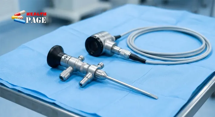
Arthroscopy Equipment Setup in an Operating Theatre
Arthroscopy equipment setup is a coordination task as much as a technical one, and it is normally handled by trained theatre staff working to hospital protocols and manufacturer instructions. In general terms, the process covers a few ideas.
The tower is positioned so the surgeon can see the monitor comfortably without twisting, and so cables can reach the patient without crossing walkways. Staff connect and check the camera, light source and monitor, then confirm that the irrigation tubing, shaver handpiece and foot pedals are present and working. Electrical connections are checked and cables are routed so they do not trail across the sterile field or become trip hazards. The working area is kept clear so that the sterile team has room to move.
Preparation also includes the sterile accessories: the arthroscope itself, sterile drapes for the camera head and cables, and any single-use or reprocessed instruments needed. Professional practice documents such as the CDC guidance on disinfection of healthcare equipment stress that device-specific instructions from the manufacturer should always guide cleaning and reprocessing.
Equipment checks usually happen before the patient is anaesthetised, so a faulty component can be replaced without delay. Facilities often keep backup equipment available for exactly this reason.
What Types of Surgery Use an Arthroscopy Tower?
Arthroscopic surgery equipment is used across several joints. The NHS notes that arthroscopy is commonly used on the knees, ankles, shoulders, elbows, wrists and hips. Surgeons usually reach the decision to operate only after the clinical picture has been assessed, often with imaging, and an introduction to reading a bone X-ray shows how that first step works.
The same tower can serve different joints, but the setup changes:
- Knee: among the most frequent applications, typically involving the meniscus, ligaments and cartilage. The knee is large enough to accommodate larger scopes and powered instruments.
- Shoulder: often relies heavily on fluid management and may involve more powered and energy-based tools.
- Hip: a deeper joint that generally calls for specialised instruments and positioning equipment alongside the tower.
- Ankle, elbow and wrist: smaller joints where smaller scopes and delicate handling are typical.
Surgical teams choose configurations to match the case. Many hospitals also use other advanced systems elsewhere in orthopaedics. Robotic platforms are one example, and they carry their own risk profile, which is discussed in this look at complications of robotic knee surgery. They are a separate category from the visualisation-focused arthroscopy tower.
The most important point is also the simplest: the tower supports visualisation and instrumentation. The surgeon’s skill, judgement and the clinical decision to operate are what determine outcomes.
Safety, Cleaning and Maintenance Considerations
Because arthroscopes enter sterile tissue, reprocessing is taken seriously. The CDC’s guideline notes that scopes such as arthroscopes are difficult to clean because of intricate designs, that meticulous cleaning must come before disinfection or sterilisation, and that newer models can often withstand steam sterilisation, which is preferred for devices of this type. It also acknowledges that evidence comparing sterilisation and high-level disinfection for these devices is limited. In practice, hospitals follow national regulations and the manufacturer’s written instructions for each device, and this article deliberately gives no universal protocol.
Other safety themes are more straightforward:
- Electrical safety: consoles, cables and foot pedals should be inspected and tested according to hospital biomedical engineering schedules.
- Sterile barriers: drapes over cameras and cables protect the sterile field while allowing nonsterile equipment to be used nearby.
- Fluid handling: pumps and tubing should be assembled according to instructions, and staff should monitor fluid use throughout the case.
- Preventive maintenance: light sources, cameras and monitors need scheduled servicing, and light cables and lenses should be checked for damage that reduces image quality.
Settings for pressure, flow, energy output and sterilisation cycles vary by device and procedure. Those details belong in manufacturer manuals and local protocols, not in general articles.
Frequently Asked Questions
What is the difference between an arthroscope and an arthroscopy tower?
The arthroscope is the slim optical instrument inserted into the joint. The tower is the wheeled platform of consoles and monitor that powers, processes and displays what the scope sees, along with supporting equipment.
Does every tower include a shaver and an irrigation pump?
Not necessarily. Configurations vary by manufacturer, hospital and procedure. Imaging components are the core, while fluid management and powered instruments are common but may be separate or procedure-dependent.
Can the same tower be used for knee and shoulder surgery?
Often yes, since the imaging chain is similar. What changes is the choice of scope, instruments, fluid management and positioning equipment for each joint.
Is image quality only about the monitor?
No. Image quality depends on the whole chain: the arthroscope lens, light cable and source, camera sensor and processor, and the monitor. A weak or damaged link anywhere reduces what the surgeon sees.
Are arthroscopy images recorded?
Many systems can capture photos or video, which supports documentation and education. Recording is subject to hospital policy, patient consent and privacy regulations.
Should patients worry about the equipment itself?
Patients are best reassured by asking their surgical team how equipment is checked and reprocessed. This article is for general understanding and does not replace that conversation.
Conclusion
An arthroscopy tower is best understood as a coordinated system rather than a single machine. Light, optics, camera, processor and display work as one chain, supported by fluid management and powered instruments that vary by procedure. Knowing the difference between the arthroscope, the camera system and the tower makes much of the language of orthopaedic theatres easier to follow, and understanding setup and reprocessing explains why theatre teams rely so heavily on checklists.
For readers who want to keep learning, the Orthopedics section of HealthPage covers related musculoskeletal topics, and manufacturer documentation and local theatre protocols remain the authority for anything hands-on. This article would also benefit from review by a qualified orthopaedic surgeon or experienced theatre practitioner before publication.
References
- NHS. Arthroscopy. https://www.nhs.uk/conditions/arthroscopy/
- Centers for Disease Control and Prevention. Disinfection of Healthcare Equipment (Guideline for Disinfection and Sterilization in Healthcare Facilities, 2008). https://cdc.gov/infection-control/hcp/disinfection-sterilization/healthcare-equipment.html

Dr. John Ma is a dedicated Orthopaedic Surgeon with extensive experience in musculoskeletal health and surgical care. He currently serves at Royal Brisbane and Women’s Hospital, Australia, where he provides expert treatment for complex orthopaedic conditions. Passionate about advancing patient outcomes, Dr. Ma combines evidence‑based practice with compassionate care, helping individuals restore mobility and improve quality of life. Through his contributions to healthcare, he continues to support patients in achieving long‑term wellness and recovery.