
Blood Flow Through the Heart: A Step-by-Step Guide
Medical Disclaimer: The information and educational content presented in “Blood Flow Through the Heart: A Step-by-Step Guide” by Dr. Akram Chang (Cardiovascular Medicine) are intended solely for informational purposes. This content is not a substitute for professional medical advice, diagnosis, or treatment. Always seek the advice of your physician or another qualified healthcare provider with any questions you may have regarding a medical condition.
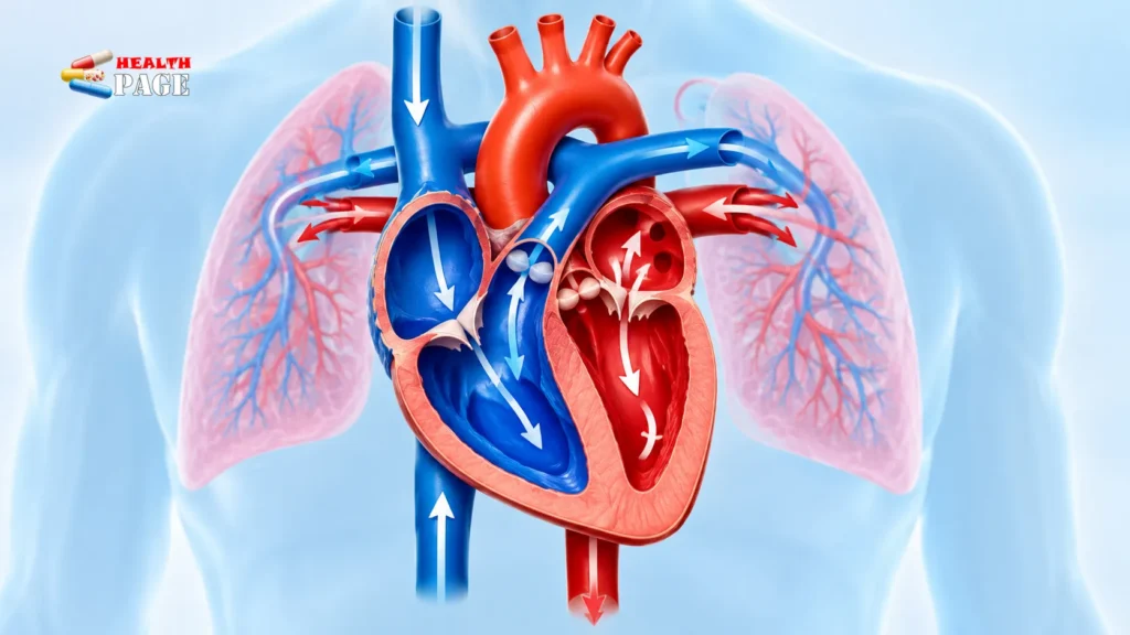
Every heartbeat moves blood along the same route, and blood flow through the heart follows that route in a fixed order. Oxygen-poor blood returns from the body to the right side, travels to the lungs, comes back as oxygen-rich blood to the left side, and is then pumped out to the rest of the body. Four valves act as one-way doors along the way, so blood keeps moving forward instead of drifting back.
That is the short answer. The rest of this guide covers what each chamber and valve does, the pathway in seven steps, and how the heart’s own blood supply differs from the blood passing through its chambers. It also explains where common misunderstandings come from. Medical terms are defined as they appear, so no background in anatomy is needed.
Heart Chambers and Valves: The Structure Behind the Route
The heart is a muscular pump roughly the size of a fist. The CDC’s overview of how the heart works describes four chambers: two atria on top and two ventricles below. A wall called the septum separates the right half from the left half. In a healthy heart, this is what keeps oxygen-poor and oxygen-rich blood apart. The atria mainly receive blood, while the ventricles do most of the pumping.
| Chamber | Main job | Receives or pumps? | Blood it handles |
|---|---|---|---|
| Right atrium | Collects blood returning from the body | Receives | Oxygen-poor |
| Right ventricle | Sends blood to the lungs | Pumps | Oxygen-poor |
| Left atrium | Collects blood returning from the lungs | Receives | Oxygen-rich |
| Left ventricle | Sends blood to the whole body | Pumps | Oxygen-rich |
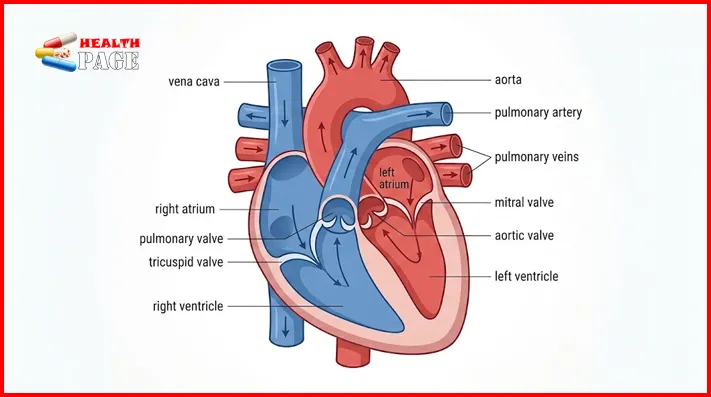
Four valves sit between the chambers and at the exits of the ventricles. Each is built from thin flaps, called cusps or leaflets, that swing open to let blood through and close to stop it slipping backward.
- Tricuspid valve: between the right atrium and right ventricle
- Pulmonary valve: between the right ventricle and the pulmonary artery
- Mitral valve: between the left atrium and left ventricle
- Aortic valve: between the left ventricle and the aorta
The tricuspid and mitral valves sit between upper and lower chambers, so they are called atrioventricular valves. The pulmonary and aortic valves guard the exits and are called semilunar valves, after their half-moon-shaped cusps. Their closing produces the familiar “lub-dub” a doctor hears through a stethoscope. The first sound comes mainly from the tricuspid and mitral valves shutting, the second from the pulmonary and aortic valves. Readers who like the house analogy of rooms, doors, pipes and wiring may enjoy this plain-language look at the structure of the human heart.
The Pathway of Blood Through the Heart, Step by Step
The sequence below follows the route described by the National Heart, Lung, and Blood Institute. It traces a single path from the body, through the heart, to the lungs, back through the heart, and out again.
- Venae cavae to the right atrium. After tissues take up oxygen, blood returns through two large veins. The superior vena cava drains the head, neck and arms, and the inferior vena cava drains the lower body. Both empty into the right atrium.
- Right atrium to right ventricle. The right atrium contracts and pushes blood through the tricuspid valve into the right ventricle.
- Right ventricle to the lungs. The right ventricle pumps blood through the pulmonary valve into the main pulmonary artery, which divides into right and left branches heading to each lung.
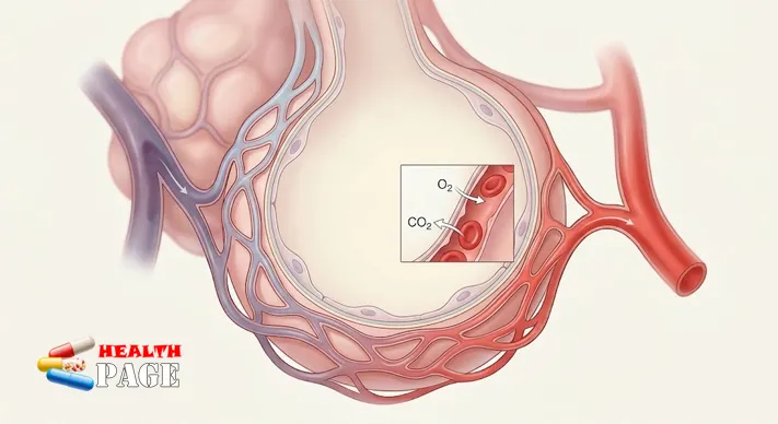
- Gas exchange in the lungs. Blood releases carbon dioxide, which is breathed out, and picks up oxygen.
- Pulmonary veins to the left atrium. Four pulmonary veins carry the newly oxygen-rich blood into the left atrium.
- Left atrium to left ventricle. Blood passes through the mitral valve, also known as the bicuspid valve, into the left ventricle.
- Left ventricle to the aorta. The left ventricle pumps blood through the aortic valve into the aorta, the body’s main artery, which branches to supply every organ.
For quick memorising, the whole pathway of blood through the heart compresses into one line:
Body → vena cava → right atrium → tricuspid valve → right ventricle → pulmonary valve → pulmonary arteries → lungs → pulmonary veins → left atrium → mitral valve → left ventricle → aortic valve → aorta → body
Step four deserves a closer look. Inside the lungs, the pulmonary arteries divide into ever smaller vessels that end in capillaries wrapped around tiny air sacs called alveoli. The walls here are extremely thin, so carbon dioxide can move out of the blood and oxygen can move in. This exchange is what turns oxygen-poor blood into oxygen-rich blood.
Although the steps are listed in order, the two sides of the heart do not take turns. The two atria contract together, then the two ventricles contract together. While the right ventricle sends one portion of blood toward the lungs, the left ventricle is sending another portion into the aorta. The list traces one path, but it is not a timeline of separate events.
The left ventricle has the thickest wall of the four chambers. It must generate the high pressure needed to drive blood through the entire body, whereas the right ventricle only has to reach the nearby lungs. Clinicians often judge how well the left ventricle is working by measuring its ejection fraction, the percentage of blood it pushes out with each contraction.
Pulmonary, Systemic and Coronary Circulation Compared
The word “circulation” covers three loops that are easy to confuse. Each one serves a different purpose.
| Loop | Route | Blood in its arteries | Purpose |
|---|---|---|---|
| Pulmonary | Right ventricle → pulmonary arteries → lungs → pulmonary veins → left atrium | Oxygen-poor | Release carbon dioxide, pick up oxygen |
| Systemic | Left ventricle → aorta → body → venae cavae → right atrium | Oxygen-rich | Deliver oxygen and nutrients, collect waste |
| Coronary | Aorta → coronary arteries → heart muscle → cardiac veins → right atrium | Oxygen-rich | Feed the heart muscle itself |
The coronary loop is the one most readers overlook. The heart’s walls are thick muscle, and they cannot take in enough oxygen from the blood flowing through the chambers, so they have a dedicated supply. According to NHLBI, the coronary arteries branch off the aorta. The left coronary artery serves the left side of the heart, while the right coronary artery supplies the right atrium and parts of both ventricles. Cardiac veins then return oxygen-poor blood from the muscle to the right atrium, rejoining the main pathway.
Among the coronary branches, the left anterior descending artery draws particular clinical attention. It feeds parts of both ventricles, and a sudden blockage there can cause a large heart attack.
Common Misconceptions About Blood Circulation Through the Heart
“Arteries carry oxygen-rich blood and veins carry oxygen-poor blood.” The real definition is about direction. Arteries carry blood away from the heart, and veins carry it toward the heart. In the systemic loop the oxygen rule happens to hold, but in the pulmonary loop it flips. The pulmonary arteries carry oxygen-poor blood to the lungs, and the pulmonary veins carry oxygen-rich blood back.
“Oxygen-poor blood is blue.” It is a darker red than oxygen-rich blood. Veins look bluish through skin because of how light penetrates and scatters in tissue, and diagrams use blue simply as a colour code.
“The heart is nourished by the blood it pumps.” Blood flowing through the chambers does not feed the heart wall. That job belongs to the coronary arteries, which is why a narrowed coronary artery can damage the heart even when the chambers are full of oxygen-rich blood.
When Valves or Vessels Interfere With Normal Flow
Normal blood flow through the heart depends on valves that open fully and close tightly, and on coronary arteries that stay open. This section describes general mechanisms only. It cannot tell anyone whether they have a heart condition.
NHLBI describes heart valve diseases as problems in which a valve leaks (regurgitation), is too narrow (stenosis), or lacks a proper opening (atresia). A leaking valve lets some blood move backward, and a narrowed one forces the chamber to push harder. Some people notice nothing at first. Others develop fatigue, breathlessness, chest pain, faintness, or a fluttering or racing heartbeat. When valve problems are not diagnosed and treated, NHLBI notes they can lead to complications including heart failure, a condition in which the heart cannot pump enough blood to meet the body’s needs.
Coronary supply can be disrupted too. When a coronary artery narrows, the muscle it feeds receives less oxygen than it needs. NHLBI lists angina, heart attack and coronary heart disease among the conditions that affect these arteries. Because the muscle powers every chamber, a reduced coronary supply can weaken the heart’s pumping even when the valves are normal.
Sudden chest pain or pressure, severe difficulty breathing, or collapse can signal an emergency. Call your local emergency number rather than waiting to see whether the symptoms pass. For ongoing symptoms such as unexplained tiredness or breathlessness, a clinician is the right person to ask.
Frequently Asked Questions
How blood flows through the heart: is there an easy way to remember the order?
Two classroom mnemonics help. “Try Pulling My Aorta” lists the valves in the order blood meets them: tricuspid, pulmonary, mitral, aortic. “LAB RAT” pairs the left atrium with the bicuspid valve and the right atrium with the tricuspid valve. A general rule also works: the right side sends blood to the lungs, the left side sends it to the body, and an atrium always comes before its ventricle.
Does oxygen-poor blood ever mix with oxygen-rich blood in a healthy heart?
No. The septum keeps the two sides separate. Some people are born with an opening in the septum or another structural difference, known as a congenital heart defect, that can allow blood to mix. These conditions are diagnosed and managed by specialists.
What coordinates the timing of all this?
A small cluster of cells called the sinoatrial node starts each beat with an electrical signal. The signal spreads across the atria, pauses briefly at a relay point called the atrioventricular node, then travels to the ventricles. That short delay lets the atria finish emptying before the ventricles squeeze.
Can healthy habits protect blood circulation?
They support the heart and blood vessels, but they cannot guarantee that a circulatory condition will never develop, because age, genetics and other factors also play a part. Not smoking, regular physical activity and a balanced diet are the habits most often recommended for heart health. For a cardiologist’s view on heart-healthy eating, there is a dedicated guide.
Why Normal Circulation Matters
A heartbeat is really two coordinated pumps working side by side. The right pump sends oxygen-poor blood to the lungs, and the left pump sends oxygen-rich blood to every tissue, including the heart’s own muscle through the coronary arteries. Valves keep that traffic moving one way, and the septum keeps the two streams separate. Once you can say the sequence aloud, blood flow through the heart stops being a list to memorise and becomes a loop you can reason about.
A practical next step is to sketch a blank heart from memory, label the chambers and valves, then check it against the diagram above. Anyone concerned about their own heart health should speak with a doctor rather than rely on a general guide. For related topics, HealthPage’s cardiology guides cover heart conditions, symptoms and tests in more depth.
References
- National Heart, Lung, and Blood Institute. “How Blood Flows through the Heart.” https://www.nhlbi.nih.gov/health/heart/blood-flow
- Centers for Disease Control and Prevention. “How the Heart Works.” https://www.cdc.gov/heart-defects/how-the-heart-works/index.html
- National Heart, Lung, and Blood Institute. “What Are Heart Valve Diseases?” https://www.nhlbi.nih.gov/health/heart-valve-diseases

Dr. Akram Chang is a veteran Cardiac Specialist with extensive clinical experience serving at the historic
Gulab Devi Teaching Hospital in Lahore.
Specializing in clinical cardiology, myocardial preservation, and preventative cardiovascular health, he dedicates his medical career to both critical inpatient care and global public health literacy.
As a core panelist on the HealthPage Medical Review Board,
Dr. Chang provides rigorous clinical oversight, expert fact-checking, and medical verification for our thoracic and circulatory care resources. His analytical oversight ensures that complex hospital diagnostics are translated into safe, actionable, and strictly evidence-based lifestyle habits.
✉ Professional Inquiries:
[email protected]