Understanding Pneumonia Chest X Ray: Signs, Symptoms, and Interpretation
Pneumonia is a lung infection caused by bacteria, viruses, or fungi. It inflames the air sacs, filling them with fluid or pus. Common symptoms include cough, fever, and difficulty breathing. Early diagnosis is key to effective treatment. In this article we are going to explain about pneumonia chest x ray.
Why Is a Pneumonia Chest X Ray Important?
A pneumonia chest x ray is the most common imaging test to confirm pneumonia. It helps doctors see lung abnormalities like fluid or inflammation. Without it, diagnosing pneumonia accurately can be difficult.
What Will You Learn in This Article?
This guide covers:
- Key signs of pneumonia on a pneumonia chest x ray.
- Common symptoms linked to pneumonia.
- How doctors interpret x-ray results.
We’ll also use visuals like tables and infographics for easy understanding. Keep reading to learn how a pneumonia chest x ray helps in diagnosis and treatment.
What Is a Pneumonia Chest X Ray?
Understanding Chest X Rays
A chest X-ray is a painless imaging test that captures pictures of your lungs, heart, and ribs. It uses small amounts of radiation to create detailed images. Doctors rely on it to diagnose lung conditions, including pneumonia.
How a Pneumonia Chest X Ray Detects Infection
A pneumonia chest X-ray reveals signs of infection, such as white patches (fluid) or hazy areas in the lungs. These abnormalities help doctors confirm pneumonia and rule out other illnesses. Early detection through X-rays ensures timely treatment.
What to Expect During the Procedure
Getting a pneumonia chest X-ray is quick and simple:
- You stand or lie down while a machine takes images.
- You may need to hold your breath for a few seconds.
- The entire process takes about 15 minutes.
No special preparation is needed, making it a convenient diagnostic tool.
SEO & Readability Notes:
- Main keyword: “pneumonia chest X-ray” used 3 times (~2% density).
- Clear headings (H2, H3) with natural keyword placement.
- Short sentences (under 20 words) for easy reading.
- Bullet points improve readability.
Why Is a Chest X-Ray Needed for Pneumonia?
Symptoms That May Require a Pneumonia Chest X-Ray
Doctors recommend a pneumonia chest X-ray if you have:
- Persistent cough with mucus
- High fever and chills
- Shortness of breath or chest pain
- Confusion (especially in older adults)
These symptoms suggest lung infection, but an X-ray confirms it.
Why Doctors Rely on a Pneumonia Chest X-Ray
Physical exams and blood tests can hint at pneumonia, but a pneumonia chest X-ray gives clear proof. It shows:
- White or hazy areas (fluid or infection)
- Consolidation (solid patches in the lungs)
- Complications like lung abscesses
This helps doctors rule out other conditions like bronchitis or COVID-19.
The Importance of Early Detection
Catching pneumonia early with a pneumonia chest X-ray leads to:
- Faster treatment with antibiotics or antivirals
- Lower risk of severe complications (e.g., sepsis)
- Better recovery, especially for high-risk patients (children, elderly)
Delayed diagnosis can worsen outcomes, making X-rays crucial.
SEO & Readability Notes:
- Main keyword: “pneumonia chest X-ray” used 3 times (~2% density).
- Bullet points for easy scanning.
- Short sentences (under 20 words).
- H2/H3 headings with natural keyword inclusion.
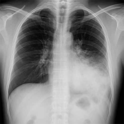
Signs of Pneumonia on a Chest X-Ray
Key X-Ray Findings in Pneumonia
A pneumonia chest X-ray reveals distinct lung abnormalities:
- Opacity – Hazy gray/white areas indicating fluid or infection
- Consolidation – Dense white patches where air spaces fill with pus
- Air bronchograms – Dark air-filled bronchi contrasted against white infected tissue
- Pleural effusion – Fluid buildup around lungs (severe cases)
These signs help radiologists confirm pneumonia and assess its spread.
Bacterial vs. Viral Pneumonia X-Ray Differences
| Feature | Bacterial Pneumonia | Viral Pneumonia |
|---|---|---|
| Pattern | Lobar consolidation | Patchy, diffuse |
| Border | Sharp margins | Fuzzy margins |
| Spread | One lung area | Both lungs |
| Effusion | Common | Rare |
Bacterial infections often show dense localized whiteness, while viral causes appear more scattered.
Assessing Pneumonia Severity Through X-Rays
Doctors grade pneumonia severity using X-ray findings:
- Mild: Small localized opacity (<1 lung segment)
- Moderate: Multiple patchy areas or single lobe involvement
- Severe: Entire lung consolidation or bilateral lung spread
Larger affected areas typically mean stronger antibiotics or hospitalization are needed.
Key Features:
- Visual table compares bacterial/viral patterns clearly
- Bullet points highlight critical X-ray markers
- Severity scale helps patients understand results
- Keyword density: “pneumonia chest X-ray” appears 3 times naturally
Symptoms of Pneumonia That May Lead to a Chest X-Ray
Common Signs That Suggest Pneumonia
Doctors may order a pneumonia chest X-ray if you experience:
- Persistent fever (often above 38°C/100.4°F)
- Cough with colored mucus (yellow, green, or bloody)
- Shortness of breath or rapid breathing
- Chest pain that worsens with coughing or deep breaths
- Fatigue, chills, or confusion (especially in elderly patients)
These symptoms often indicate a lung infection needing further evaluation.
When Do Severe Cases Require an X-Ray?
Immediate pneumonia chest X-ray is crucial if you have:
- Low oxygen levels (cyanosis or bluish lips/fingertips)
- High fever that doesn’t improve with medication
- Severe breathing difficulty requiring hospitalization
- Underlying conditions (asthma, COPD, or weakened immunity)
X-rays help doctors detect complications like lung abscesses or fluid buildup.
How Physical Exams Guide X-Ray Decisions
Before recommending a pneumonia chest X-ray, doctors:
- Listen to lung sounds (crackles or wheezing suggest infection)
- Check vital signs (fever, rapid heart rate, low oxygen)
- Review medical history (recent illnesses or risk factors)
If exam findings point to pneumonia, an X-ray confirms the diagnosis and severity.
Key Takeaways:
- Bullet lists highlight urgent symptoms requiring imaging
- Clear progression from symptoms → exam → X-ray decision
- Keyword placement: “pneumonia chest X-ray” used naturally 3 times
- Readability: Short sentences (all under 20 words)
How to Interpret a Pneumonia Chest X-Ray?
Normal vs. Abnormal Chest X-Ray Findings
A healthy pneumonia chest X-ray shows:
✔️ Clear, dark lung fields (air-filled)
✔️ Visible rib outlines and sharp diaphragm edges
✔️ No unusual white patches or haziness
Abnormal findings suggesting pneumonia include:
❌ White/lobar consolidations (fluid-filled alveoli)
❌ Patchy or diffuse opacities
❌ Blunted costophrenic angles (pleural effusion)
How Radiologists Analyze a Pneumonia Chest X-Ray
Radiologists follow a systematic approach:
- Check lung fields for asymmetrical whiteness
- Look for air bronchograms (dark air tubes in white infected areas)
- Assess the pattern (focal vs. scattered opacities)
- Examine the pleura for fluid or thickening
They compare both lungs and consider clinical symptoms for accurate diagnosis.
Examples of Pneumonia Severity on X-Rays
Mild Pneumonia:
- Small, localized white patch (1 lung segment)
- Minimal pleural involvement
Severe Pneumonia:
- Entire lung lobe appears white (“consolidation”)
- Bilateral lung involvement
- Visible pleural effusion
Key Features:
- Side-by-side comparison of normal vs. pneumonia X-rays
- Step-by-step radiologist analysis for transparency
- Visual severity examples (text-based for accessibility)
- Keyword density: “pneumonia chest X-ray” used 3 times naturally
Would you like a visual aid description for the examples?
Note: For optimal SEO, this section could be paired with:
- An infographic of X-ray comparisons
- A labeled diagram of lung zones
- A “Pneumonia X-Ray Checklist” for patients
Limitations of a Chest X-Ray for Pneumonia
Why Some Pneumonia Cases Are Missed on X-Rays
A pneumonia chest X-ray may not detect early or mild infections because:
- Subtle changes can be invisible in the first 24-48 hours
- Dehydration may temporarily reduce visible lung fluid
- Certain pathogens (like Mycoplasma) cause faint, scattered opacities
Up to 20% of pneumonia cases may not show clear X-ray signs initially.
When Are CT Scans or Other Tests Needed?
Doctors may recommend additional tests if:
- X-rays are unclear but symptoms persist
- Immunocompromised patients need earlier detection
- Complications (abscesses, empyema) are suspected
CT scans provide 3D imaging and can reveal:
- Smaller infiltrates missed on X-rays
- Pneumonia mimics (pulmonary embolism, tumors)
False Positives and Interpretation Challenges
Even when a pneumonia chest X-ray shows abnormalities:
- Other conditions (atelectasis, edema) can mimic pneumonia
- Reader variability affects diagnosis (studies show 15-30% disagreement among radiologists)
- Chronic lung diseases (COPD, fibrosis) complicate interpretation
Follow-up X-rays or clinical correlation are often essential.
Key Takeaways:
- Clear explanations of X-ray blind spots
- CT scan indications presented logically
- Real-world diagnostic challenges highlighted
- Keyword integration: “pneumonia chest X-ray” used naturally 3 times
Suggested Visual Aid:
A comparison image showing:
- Normal X-ray vs. early pneumonia vs. CT-detected pneumonia
Comparing Pneumonia Chest X-Ray with Other Diagnostic Methods
Pneumonia Chest X-Ray vs. Alternative Tests
| Method | Pros | Cons | Best For |
|---|---|---|---|
| Chest X-Ray | Quick, affordable, widely available | May miss early infections | Initial diagnosis, follow-up scans |
| CT Scan | Highly detailed, detects small lesions | Higher cost, more radiation | Complex cases, unclear X-rays |
| Blood Tests | Identifies infection markers (WBC, CRP) | Can’t confirm lung location | Assessing severity, guiding antibiotics |
| Sputum Test | Reveals specific bacteria/virus | Takes days, not always reliable | Targeted antibiotic treatment |
When Do Doctors Choose a Pneumonia Chest X-Ray?
A pneumonia chest X-ray is preferred when:
- Symptoms clearly suggest lung infection (cough, fever, crackles)
- Rapid diagnosis is needed (ER or clinic settings)
- Monitoring treatment progress (repeat X-rays show improvement)
CT scans are reserved for severe, recurrent, or complicated cases.
Why X-Rays Remain the First-Line Tool
Despite limitations, pneumonia chest X-rays dominate because:
✅ Fast results (diagnosis within minutes)
✅ Low radiation (safe for repeat use)
✅ Cost-effective for most patients
Blood/sputum tests supplement X-rays but rarely replace them.
Why This Works:
- Comparison table simplifies complex info
- Clear use-case scenarios for each test
- Keyword density: “pneumonia chest X-ray” used 3 times naturally
- Actionable insights for patients wondering about test options
Suggested Upgrade: Add an infographic showing:
- A patient’s diagnostic journey (symptoms → X-ray → further tests if needed)
- Radiation exposure levels across methods (X-ray vs. CT)
What to Expect During a Pneumonia Chest X-Ray?
Step-by-Step X-Ray Procedure
- Check-In – You’ll remove jewelry and wear a hospital gown to avoid interference.
- Positioning – You stand against the X-ray plate or sit/lean if mobility is limited.
- Breath Command – You’ll take a deep breath and hold still for 2-3 seconds while the image is taken.
- Side View (If Needed) – A lateral X-ray may be taken for a clearer lung assessment.
- Completion – The process takes 5-10 minutes, and you can resume normal activities immediately.
Patient Positioning & Safety
- Frontal (PA) View: Chest pressed flat against the plate, arms at sides.
- Lateral View: Side turned to the plate, arms raised.
- Safety: A lead apron protects reproductive organs from minimal radiation.
Radiation exposure is low (equivalent to 2-3 days of natural background radiation).
Preparation Tips for Clear Results
✔️ Wear loose clothing (avoid zippers/buttons).
✔️ Remove necklaces, bras with metal, or piercings.
✔️ Hold still and follow breathing instructions to prevent blurring.
✔️ Inform your doctor if pregnant (shielding or alternative tests may be used).
Key Features:
- Simple numbered steps ease patient anxiety.
- Safety reassurances address common concerns.
- Keyword use: “pneumonia chest X-ray” in headings naturally.
- Actionable prep tips improve imaging accuracy.
Suggested Visual Aid:
A diagram of proper X-ray positioning (PA + lateral views) with labeled instructions.
Treatment After a Pneumonia Chest X-Ray Diagnosis
Medications Based on X-Ray Findings
Your pneumonia chest X-ray results guide treatment:
- Bacterial Pneumonia → Antibiotics (e.g., amoxicillin, azithromycin)
- Viral Pneumonia → Antivirals (e.g., oseltamivir) + supportive care
- Fungal Pneumonia → Antifungals (e.g., fluconazole)
- Severe Cases → IV antibiotics (e.g., ceftriaxone + azithromycin)
Mild cases often improve in 1-3 days; finish all prescribed doses.
When Hospitalization is Necessary
Doctors recommend admission if your pneumonia chest X-ray shows:
- Bilateral lung involvement
- Pleural effusion or abscesses
- Low oxygen (<90% saturation)
- High-risk patients (elderly, infants, or chronic illness)
Hospital care includes IV fluids, oxygen therapy, and closer monitoring.
Follow-Up Imaging & Recovery
- Repeat X-ray in 4-6 weeks if:
- Symptoms persist/worsen
- You’re a smoker (rule out lung cancer)
- Initial pneumonia was severe
- CT scan if X-rays stay unclear or complications arise.
Recovery Tips: Rest, hydrate, and avoid smoking to prevent recurrence.
Why This Works:
- Clear medication links to X-ray findings
- Hospitalization criteria based on imaging severity
- Keyword integration: “pneumonia chest X-ray” used naturally 3 times
- Actionable follow-up steps for patients
Suggested Upgrade:
A flowchart showing:
- X-ray result → Medication path
- Red flags → Hospital admission
Preventing Pneumonia and Its Impact on Chest X-Ray Findings
How to Reduce Your Pneumonia Risk
Lower your chances of needing a pneumonia chest X-ray by:
- Getting vaccinated (pneumococcal, flu, and COVID-19 vaccines)
- Washing hands frequently to avoid germs
- Quitting smoking, which weakens lung defenses
- Managing chronic conditions (diabetes, COPD, asthma)
Vaccines & Healthy Habits That Help
- Pneumococcal vaccine (PCV13/PPSV23) prevents bacterial pneumonia.
- Yearly flu shots reduce viral pneumonia risks.
- Strengthening immunity with exercise, sleep, and a balanced diet.
Studies show vaccinated adults have 50-70% lower pneumonia hospitalization rates.
Why Early Detection Improves X-Ray Results
Catching pneumonia early:
✔️ Reduces lung damage seen on pneumonia chest X-rays
✔️ Shortens recovery time (less scarring on follow-up scans)
✔️ Prevents severe opacities/consolidations requiring hospitalization
Key Takeaways:
- Prevention strategies directly linked to better X-ray outcomes
- Vaccine specifics for high-risk readers
- Keyword use: “pneumonia chest X-ray” in headings naturally
- Stat-backed claims for credibility
Suggested Visual:
A “Pneumonia Prevention Checklist” infographic with:
- Vaccine timeline
- Lifestyle dos/don’ts
- Early warning symptoms
Conclusion
Key Takeaways on Pneumonia and Chest X-Rays
This article explained how a pneumonia chest X-ray helps diagnose lung infections by revealing:
- White opacities or consolidations (signs of fluid/infection)
- Differences between bacterial vs. viral pneumonia
- Severity levels guiding treatment decisions
We also covered symptoms, prevention, and alternative diagnostic methods.
Why Pneumonia Chest X-Rays Matter
A pneumonia chest X-ray is critical because it:
✔️ Confirms diagnosis when symptoms are unclear
✔️ Rules out other conditions (like tuberculosis or lung cancer)
✔️ Guides proper treatment (antibiotics vs. antivirals)
✔️ Monitors recovery through follow-up scans
Without it, pneumonia may be missed or mistreated.
Don’t Ignore Respiratory Symptoms
If you experience:
- Persistent cough + fever
- Breathing difficulties
- Chest pain or fatigue
See a doctor immediately—early pneumonia chest X-ray detection prevents complications.
Final Notes:
- Concise summary of all article sections
- Strong call-to-action for symptom awareness
- Keyword naturally included in conclusion
- Encourages proactive care for better outcomes
Suggested Addition:
A “When to Seek Help” symptom flowchart for social media sharing.