
7 Powerful and Amazing Benefits of doppler ultrasound for Early Disease Detection
Many serious health problems start quietly.
They grow for months or years before symptoms appear.
That is where doppler ultrasound can make a life‑saving difference.
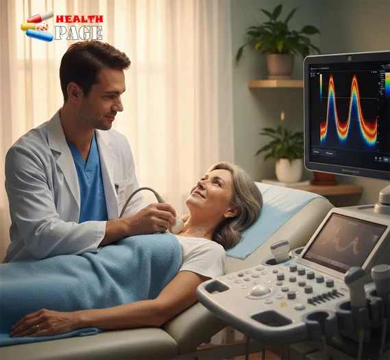
This noninvasive scan shows how blood moves through your arteries and veins.
It helps doctors see trouble before it becomes an emergency.
If you have ever wondered what is a doppler ultrasound, you are not alone.
Many people feel nervous before any test.
Knowing how it works and why it matters can ease that worry.
It can also help you ask better questions and protect your health.
In this article, you will learn seven key benefits of this test.
You will see how it helps find disease early, guide treatment, and give peace of mind.
Understanding the Basics: what is a doppler ultrasound? 🩻
A standard ultrasound uses sound waves to create pictures of organs.
A doppler ultrasound goes one step further.
It also measures how blood moves through your vessels.
Here is how it works in simple terms:
- A small handheld device (transducer) moves over your skin.
- It sends high‑frequency sound waves into your body.
- These waves bounce off moving red blood cells.
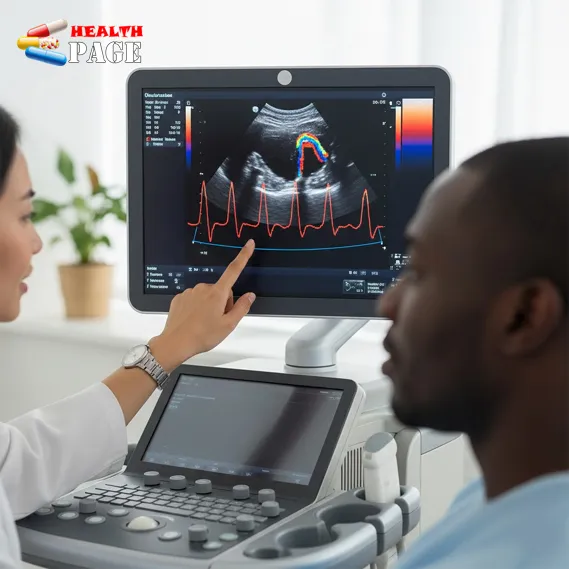
- A computer turns those echoes into images and color maps.
- Your doctor sees both structure and blood flow in real time.
You do not feel the sound waves.
There is no radiation.
Most exams take 20–60 minutes and you go home right after.
Doctors often use this scan to study:
- Neck arteries that supply the brain
- Leg arteries and veins
- Heart and major vessels
- Blood flow in organs like kidneys or liver
- Blood flow in a baby during pregnancy
1. Finds Blood Clots Before They Turn Dangerous 🧬
Blood clots in the legs, called deep vein thrombosis (DVT), can be dangerous.
A clot can break loose and travel to the lungs.
This can cause a life‑threatening blockage called pulmonary embolism.
A vascular ultrasound can spot these clots early.
It shows:
- Where the clot sits in the vein
- Whether blood flow is blocked or only slowed
- How large and how long the clot might be
Real‑life example
A person notices one calf looks swollen and feels warm.
Walking hurts more than usual.
Their doctor orders a leg doppler ultrasound the same day.
The scan reveals a fresh clot, and treatment starts at once.
This fast action may prevent a serious emergency.
2. Reveals Narrowed Arteries Before a Stroke or Heart Attack ⚠️
Plaque can build up inside your arteries over time.
It often causes no symptoms at first.
Yet it increases the risk of stroke and heart attack.
A doppler ultrasound can show:
- If arteries in the neck (carotid arteries) are narrowed
- How fast blood flows through those narrowed spots
- Whether plaque looks smooth or irregular
When blood flow speeds up through a tight spot, the machine detects it.
This gives your doctor an early warning sign.
With that information, your care team can:
- Adjust cholesterol or blood pressure medicines
- Recommend lifestyle changes
- Decide if a procedure to open the artery is needed
Real‑life example
Someone has high blood pressure and a family history of stroke.
They feel healthy and have no warning signs.
A carotid doppler ultrasound shows a moderate narrowing on one side.
Their doctor intensifies treatment, helping reduce future stroke risk.
3. Monitors Blood Flow in Pregnancy and Protects Baby’s Health 🤰
During pregnancy, blood must flow smoothly to the placenta and baby.
Sometimes that flow is reduced.
This can affect growth and oxygen supply.
In high‑risk pregnancies, a doppler ultrasound helps monitor:
- Blood flow in the umbilical cord
- Circulation in the baby’s brain and major vessels
- Blood flow in the mother’s uterine arteries
This test does not replace a normal pregnancy ultrasound.
Instead, it adds extra detail about circulation.
Doctors use this information to:
- Decide how closely to monitor the pregnancy
- Adjust timing of delivery if needed
- Plan care in case the baby needs help at birth
Short example
A pregnant person with high blood pressure has regular scans.
One doppler study shows reduced blood flow in the cord.
The team increases monitoring and plans an earlier delivery.
The baby arrives safely with special support ready.
4. Tracks Peripheral Artery Disease Before It Limits Your Life 🚶♂️
Peripheral artery disease (PAD) affects blood flow to the legs and feet.
It can cause pain when walking, poor wound healing, and infection.
Many people think leg pain is “just aging.”
In reality, it can be a sign of blocked arteries.
A doppler ultrasound of leg arteries can:
- Show where arteries are narrowed or blocked
- Measure how severe the blockage is
- Guide choices about medicine, exercise, or surgery
This is essential for early detection.
Treating PAD early helps prevent ulcers, infections, and even amputation.
Simple comparison table
Here is how a standard ultrasound differs from a doppler study:
| Feature | Standard Ultrasound | Doppler Ultrasound |
|---|---|---|
| Shows blood flow | No | Yes, color and wave patterns |
| Measures speed and direction | No | Yes |
| Detects narrowed vessels | Limited | Yes, often very precisely |
| Typical use | Organs, pregnancy, soft tissues | Arteries, veins, and circulation |
| Helps guide vascular treatment | Sometimes | Often essential |
A trusted RadiologyInfo vascular ultrasound resource explains these tests in more technical detail:
https://www.radiologyinfo.org/en/info/vascularUS
5. Helps Prevent Serious Complications After Surgery 🏥
After vascular or heart surgery, blood flow can change.
A bypass graft or stent may narrow or clot.
Catching these issues early is critical.
A doppler ultrasound is often used to:
- Check blood flow through new bypass grafts
- Monitor stents in arteries of the legs or neck
- Watch transplanted organs, such as kidneys, for supply problems
Because the test is repeatable and noninvasive, it is ideal for follow‑up.
Your doctor can compare today’s scan with older ones.
Small changes in flow can be addressed before they become major issues.
Short example
A patient has a bypass in a leg artery.
Months later, they return for routine imaging.
The doppler study shows slightly reduced flow through the graft.
Their team adjusts treatment and avoids graft failure.
6. Offers a Safer Option for People Who Cannot Have Contrast Dye 🌱
Some imaging tests use contrast dye.
That dye can stress the kidneys or cause allergic reactions.
People with kidney disease or strong allergies may need other options.
A doppler ultrasound often avoids these concerns.
Most exams use no contrast at all.
When contrast is used, it is usually a special ultrasound contrast.
This type is different from CT or MRI dye.
As a result, this test can be especially helpful if you:
- Have chronic kidney disease
- Are pregnant
- Have had a past reaction to contrast dye
- Need repeated imaging over many years
It lets doctors still see how blood moves through your body.
You get important answers with lower risk.
7. Gives Clear Guidance Without Radiation or Injections 🌟
Many people worry about radiation exposure.
They also fear needles and injections.
A doppler ultrasound offers several comforts:
- No ionizing radiation
- Often no needles or injections
- Usually done in a quiet, low‑stress setting
- You can often return to normal activity right away
From a medical perspective, this matters too.
Because the test is safe, doctors can repeat it over time.
They can watch how a condition changes and adjust care.
Emotional benefit
There is also a major emotional advantage.
When you understand the images and results, fear often decreases.
You feel more involved in your care.
Many patients say that seeing blood flow on the screen makes the problem feel more real and manageable.
When Might Your Doctor Recommend a Doppler Ultrasound? 🔍
Your doctor might suggest this test if you have:
- Leg pain when walking that eases with rest
- Swelling, warmth, or redness in a leg
- A whooshing sound in the neck heard with a stethoscope
- Nonhealing wounds on feet or toes
- Sudden changes in arm or leg color or temperature
- High‑risk pregnancy or reduced fetal movements
In some cases, it is part of routine follow‑up.
In others, it is an urgent test to rule out a dangerous clot or blockage.
How to Prepare and What to Expect 😊
Preparation depends on the body area.
Your care team will give specific instructions.
Often, you can expect:
- No special diet for leg or neck vessels
- Fasting for several hours for abdominal vessels
- Loose clothing for easy access to the area
During the test:
- You lie on a table, usually on your back or side
- Gel is placed on your skin to help sound waves travel
- The transducer glides over your skin with gentle pressure
- You may hear whooshing sounds of blood flow
- You may be asked to change positions or hold your breath briefly
Most people describe the test as painless.
A bit of pressure from the probe can feel uncomfortable if the area is tender.
Results often go to your referring doctor, who will explain what they mean.
FAQs About Doppler Ultrasound ❓
1. Is a doppler ultrasound painful?
Most people feel no pain.
You may feel mild pressure from the probe on your skin.
If an area is already sore, that pressure can feel uncomfortable.
The exam does not involve needles in most cases.
2. Is it safe to have this test while pregnant?
Yes, when medically needed and done by trained professionals.
It uses sound waves, not radiation.
Doctors balance the benefits and only order it when helpful for mother or baby.
3. How long does the test usually take?
Most exams take 20 to 60 minutes.
Time varies with the body area and how complex your circulation is.
You can usually go home or back to normal activity afterward.
4. Will I get results right away?
Sometimes, yes.
If a radiologist or vascular specialist is present, they may discuss impressions.
Often, a formal report goes to your main doctor.
They will review results with you and explain next steps.
5. Does insurance usually cover doppler ultrasound?
In many regions, yes, when the test is medically necessary.
Coverage depends on your plan and local rules.
It is wise to check with your insurer or clinic billing office in advance.
6. Can this test replace all other imaging?
Not always.
It is excellent for many blood flow questions.
But CT, MRI, or angiography may still be needed in some situations.
Your doctor chooses the best test or combination for your case.
Conclusion: Using Doppler Ultrasound to Protect Your Future Health 💖
Early detection can change the course of disease.
That is where doppler ultrasound truly shines.
It can:
- Find blood clots before they cause emergencies
- Reveal narrowed arteries before stroke or heart attack
- Protect babies in high‑risk pregnancies
- Track peripheral artery disease and guide treatment
- Prevent complications after vascular and heart surgery
- Offer safer imaging for people who cannot have dye
- Deliver clear answers without radiation or injections
If you have risk factors like smoking, diabetes, high blood pressure, or leg pain, talk with your doctor.
Ask whether a circulation study, including a doppler ultrasound, might be helpful.
You deserve clear information and timely care.
Understanding what is a doppler ultrasound and how it works is one step toward that goal.
With the right tests at the right time, you and your care team can act early, protect vital organs, and support a healthier future. 🌈